
Neuroradiology
Neuroradiology is an imaging subspecialty focused on diagnosing conditions of the brain, neck and spine.
At Diagnostic Radiology, P.C. (DRPC), we offer a complete range of neuroradiology exams. With an array of imaging modalities, our radiologists are able to provide referring physicians multiple choices and often assist in recommending the best imaging modality for a particular concern. An accurate interpretation of these tests can identify dangerous problems early, when treatment can be most effective.
CT and MR Imaging
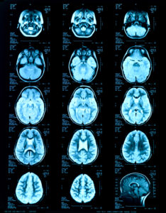 CT and MR imaging of the brain, neck and spine have vastly improved detection and diagnosis of a variety of disorders, diagnoses that can be relayed to the ordering physician who then can institute a treatment program for the various disorder that may exist. A head CT scan is commonly utilized for the initial work-up for patients with a history of trauma, severe acute-onset headache, or suspected intracranial bleeding or stroke. CT is also the modality of choice to evaluate the integrity of bony structures and can also be used to identify degenerative changes of the spine in patients who cannot undergo MRI. Brain MRI is more sensitive in diagnosing and characterizing ischemia strokes, metabolic disorders, infection and inflammatory processes and tumors. Imaging of neck can be performed with either CT or MRI, each modality has its strengths and weaknesses.
As with the head, a CT scan is often the best initial exam. Imaging of the spine is extremely common and often used to evaluate the degenerative changes as they relate to intervertebral disks and facet joints. MRI is the modality of choice because it provides the best image of the spinal canal with clear delineation of the spinal cord and nerve roots.
CT and MR Angiography (CTA and MRA)
Angiography is primarily used to diagnose vascular disorders. Often, angiography is performed to evaluate for potential narrowing and/or blockages of the carotid and vertebral arteries (arteries which supply blood to the brain). Also, these two modalities can detect abnormalities of the major draining veins in the head and neck. CTA and MRA have largely replaced catheter angiography as the imaging modality of choice because they are noninvasive in contrast to the catheter angiogram. MRA can even be performed without contrast agents, a particular medical concern in patients with diabetes, renal insufficiency, or certain allergies.
Ultrasound Imaging
The role of ultrasound in neuroradiology usually pertains to the thyroid gland or as a screening exam to evaluate the carotid and vertebral arteries. Thyroid nodules are common and ultrasound plays a critical role in work-up of thyroid nodules. Doppler ultrasound is often employed as the initial test to evaluate for potential narrowings and/or blockages of the major arteries supplying the brain. If a vascular abnormality is suspected, a CTA or MRA is commonly used to better evaluate the abnormality.
PET Imaging
PET scanning is frequently used in regards to cancer, in particular, the therapeutic response of radiation and/or chemotherapy. In the field of neuroradiology this is true of most neck cancers. PET is infrequently used in the setting of brain cancer, more commonly PET can be employed to evaluate for Alzheimer’s disease and those with a seizure disorder.
WHAT OUR PATIENTS SAY
![]()
We want to hear from you! Please submit your reviews to us here!